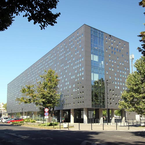
Partner 1
Wroclaw University of Science and Technology
Professor Marcin Drąg
Chemistry Faculty
Department of Chemical Biology and Bioimaging
50-370 Wroclaw, Poland
+48 71 320 4526
http://www.bioorganic.ch.pwr.wroc.pl/
index.php/Marcin_Drąg
Description
The Imaging Laboratory created by Prof. Marcin Drąg at the Department of Biological Chemistry and Bioimaging of the Wroclaw University of Science and Technology has a mass cytometer, currently the most modern device for analyzing and diagnosing cell samples as well as for advanced proteomics.
Mass cytometry is an analytical technique that has revolutionized modern medical diagnostics. The mass cytometer is therefore used to analyze samples, mainly cells, which in this process are marked with stable transition metal isotopes, mainly lanthanides. The device can examine any type of cell, e.g. cells from cancerous tumors or leukemia, blood cells or even cells from other organisms (parasites or bacteria).
The cytometer will be used primarily to study proteolytic enzymes (proteases). Proteases are specialized proteins that break down peptide bonds and are able to “cut" other proteins into simpler elements – peptides and amino acids. In humans, proteases are a group of about 700 enzymes and are involved not only in the simple digestion of food, but are also responsible for controlling key cellular processes. Their malfunction leads to the formation of pathological conditions in the body, and consequently to the development of civilization diseases such as cancer, diabetes, hypertension and viral and bacterial infections. Studies on protease activity are very important in the early diagnosis and treatment of patients.
Prof. Drąg ‘s Imaging Laboratory in cooperation with the Laboratory of Prof. Guy Salvesen (SBP Medical Discovery Institute, La Jolla, USA) has created a completely new diagnostic method that is extremely competitive compared to those currently used in mass cytometry.
In the Bioimaging Laboratory scientists will also use a confocal microscope to determine cell parameters by fluorescence methods in live and fixed cells. The microscope also allows imaging of living cells and processes occurring within them, because it is equipped with a temperature control chamber and nozzles for regulating carbon dioxide and oxygen levels.
Laboratory and equipment
under construction
Research activities
under construction
Achievements and awards
under construction
